Lower Back Muscle Diagram ~ Low Back Pain A Guide For Coaches And Athletes On Anatomy Types And Treatment Breaking Muscle. Short of a great deal of descriptive text, the easiest way to answer this is with. Deep muscles of the lower back include: Don duff aug 20, 2019 the low back is a fairly complicated structure, so it's no wonder the majority of people—experts estimate up to 80% of us—experience pain in this area of our bodies at some point in our lives. It comprises the vertebral column (spine) and two compartments of back muscles; Back to tracking tools main page.
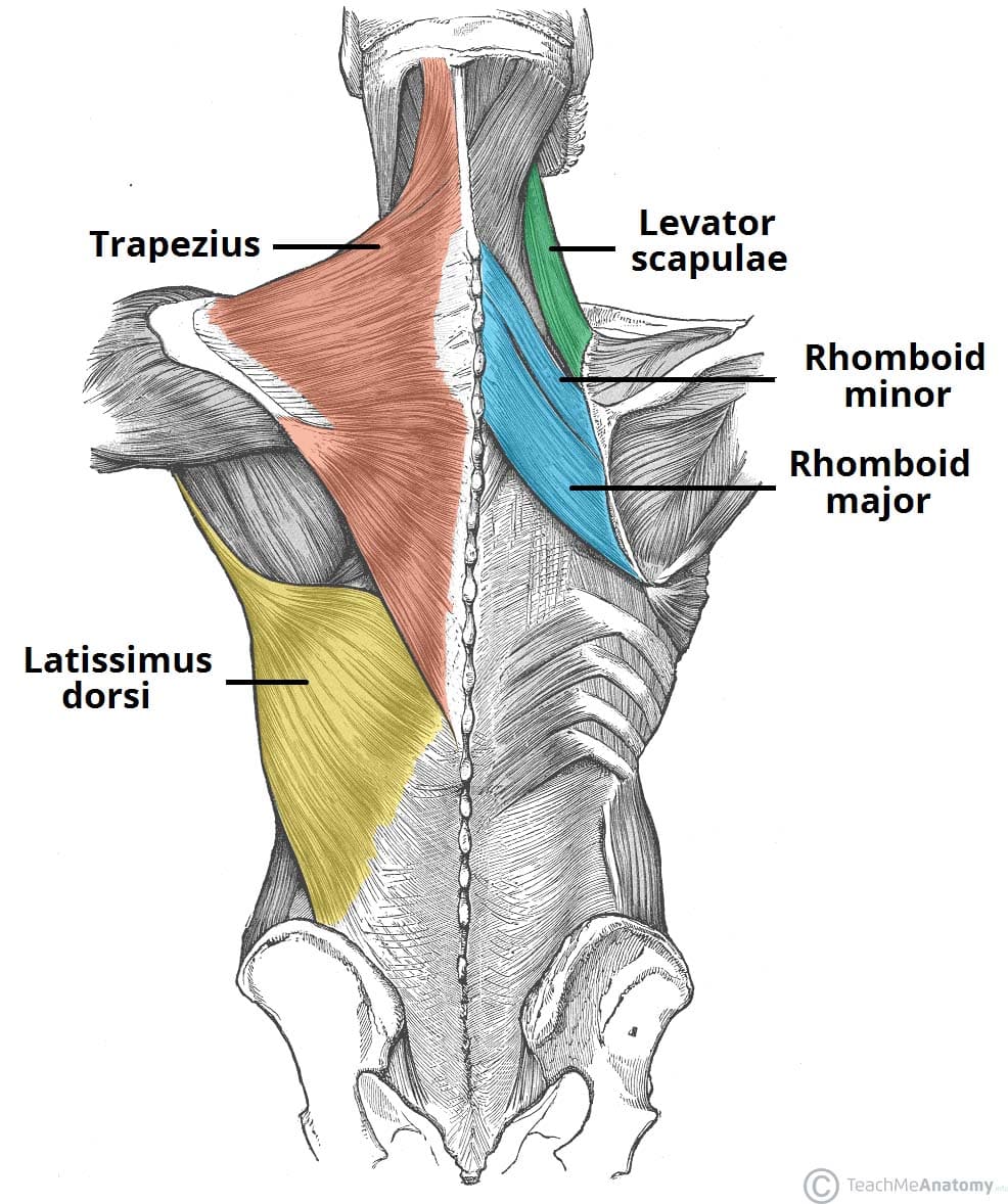
As you can see, there are also have a spine of scapula deltoid, triceps brachii, latissimus dorsi. Begin with this first one of the exercises for back pain: People with back pain people who experience headaches printing for use during doctor visits to communicate information about your symptoms quickly tracking your progress over time related tools: Lumbar muscle strain is caused when muscle fibers are abnormally stretched or torn. These structures work together to support the body, enable a range of movements, and send messages from the brain to.
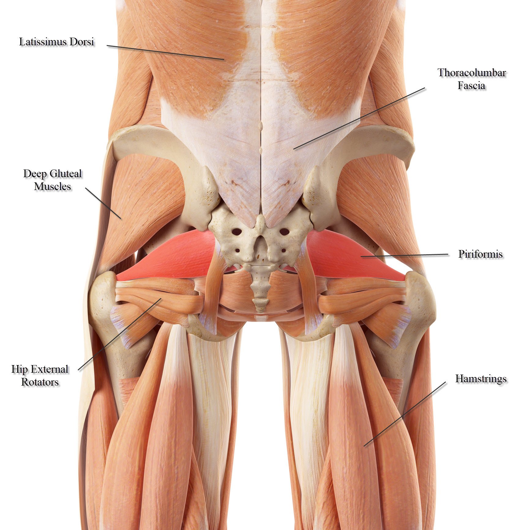
Lower Back Muscle Anatomy And Low Back Pain from ix-cdn.b2e5.com Lower back muscle diagram anatomy does degenerative disc disease affect the lower back muscle? See more ideas about muscle anatomy, anatomy, anatomy and physiology. Begin with this first one of the exercises for back pain: They help to bend the back to one side or the other. Back muscles, back muscle diagram. Pain log more pain mapping tools The bones of the pelvis and lower back work together to support the body's weight, anchor the abdominal and hip muscles, and protect the delicate vital organs of the vertebral and abdominopelvic cavities. This muscle is a major generator of lower back and hip pain, as well as being responsible for complaints of a burning sensation along the posterior superior iliac spine (psis) and sacroiliac joint.
It's a staple of the best back workouts for men , but make no mistake, it's great for back workouts for women , as well.
The bones of the pelvis and lower back work together to support the body's weight, anchor the abdominal and hip muscles, and protect the delicate vital organs of the vertebral and abdominopelvic cavities. Extrinsic and intrinsic.the back functions are many, such as to house and protect the spinal cord, hold the body and head upright, and adjust the movements of the upper and lower limbs. It's a staple of the best back workouts for men , but make no mistake, it's great for back workouts for women , as well. The four muscle groups that together make up the deep muscle group are the segmental muscles, the transversospinales, the erector spinae, and the spinotransversales. Another common cause of lower back and hip pain is disc injury. Don duff aug 20, 2019 the low back is a fairly complicated structure, so it's no wonder the majority of people—experts estimate up to 80% of us—experience pain in this area of our bodies at some point in our lives. This is a diagram of the larger and more surface muscles of the low back. The muscles of the lower back, including the erector spinae and quadratus lumborum muscles, contract to extend and laterally bend the vertebral these muscles provide posture and stability to the body by holding the vertebral column erect and adjusting the position of the body to maintain balance. Short of a great deal of descriptive text, the easiest way to answer this is with. Lumbar (lower back) muscle strains and sprains are the most common causes of low back pain. Muscle anatomy neck 12 photos of the muscle anatomy neck dog neck muscle anatomy, front neck muscle anatomy, muscle anatomy neck, muscle anatomy of neck and shoulder, neck muscle anatomy chart, human muscles, dog neck muscle anatomy, front neck muscle anatomy, muscle anatomy neck, muscle anatomy of neck and. Nerves in your lower back. Lower back and superficial muscles the muscles of the lower back help stabilize, rotate, flex, and extend the spinal column, which is a bony tower of 24 vertebrae that gives the body 13.04.2020 · related posts of muscles of the lower back and hip diagram foot muscle anatomy mri.
Neck muscle anatomy ultrasound 12 photos of the neck muscle anatomy ultrasound , human muscles. While muscles like the gluteals (in the thighs) are used any time we walk or climb a step, deep back muscles and abdominal muscles are usually not actively engaged during everyday activity. Muscles of lower back diagram in this image, you will find an occipital bone, sternocleidomastoid, trapezius, deltoid in muscles of the lower back diagram. It comprises the vertebral column (spine) and two compartments of back muscles; The back consists of the spine, spinal cord, muscles, ligaments, and nerves.
Muscles Move And Support The Spine from cloud2.spineuniverse.com Lower back and superficial muscles the muscles of the lower back help stabilize, rotate, flex, and extend the spinal column, which is a bony tower of 24 vertebrae that gives the body 13.04.2020 · related posts of muscles of the lower back and hip diagram foot muscle anatomy mri. See how exercise helps the back. Inhale as you raise your hands up to the side and interlace your fingertips together behind your head. The quadratus lumborum muscles (orange, in the image above) are found in the lower back (also called the lumbar area). See more ideas about muscle anatomy, anatomy, anatomy and physiology. Lumbar muscle strain is caused when muscle fibers are abnormally stretched or torn. Lower back muscle diagram anatomy does degenerative disc disease affect the lower back muscle? These structures work together to support the body, enable a range of movements, and send messages from the brain to.
As you can see, there are also have a spine of scapula deltoid, triceps brachii, latissimus dorsi.
The muscles that move the upper legs (thigh) there are many muscles that move the large bone of the thigh. Cure lower back pain naturally. This is a diagram of the larger and more surface muscles of the low back. The four muscle groups that together make up the deep muscle group are the segmental muscles, the transversospinales, the erector spinae, and the spinotransversales. Out of these, the cookies that are categorized as necessary are stored on your browser as they are essential for the working of basic functionalities of the website. Deadlift muscles will include knee, hip, and back extensors, which primarily include the quads, glutes, and spinal erectors. Muscles of lower back diagram in this image, you will find an occipital bone, sternocleidomastoid, trapezius, deltoid in muscles of the lower back diagram. The muscles of the lower back, including the erector spinae and quadratus lumborum muscles, contract to extend and laterally bend the vertebral these muscles provide posture and stability to the body by holding the vertebral column erect and adjusting the position of the body to maintain balance. The multifidus, a long muscle that travels nearly the entire length of the back.it helps to stabilize and rotate the lower back, and additionally takes some. Lumbar muscle strain is caused when muscle fibers are abnormally stretched or torn. Extrinsic and intrinsic.the back functions are many, such as to house and protect the spinal cord, hold the body and head upright, and adjust the movements of the upper and lower limbs. Another common cause of lower back and hip pain is disc injury. See how exercise helps the back.
This website uses cookies to improve your experience while you navigate through the website. Another common cause of lower back and hip pain is disc injury. Deep muscles of the lower back include: See how exercise helps the back. Nerves in your lower back.
Muscles Of The Back Teachmeanatomy from teachmeanatomy.info Lower back muscle diagram anatomy does degenerative disc disease affect the lower back muscle? The vertebral column of the lower back includes the five lumbar vertebrae, the sacrum, and the coccyx. They help to bend the back to one side or the other. Deep muscles of the lower back include: The back is the body region between the neck and the gluteal regions. The extrinsic back muscles, which lie most superficially on the back. This muscle is a major generator of lower back and hip pain, as well as being responsible for complaints of a burning sensation along the posterior superior iliac spine (psis) and sacroiliac joint. See more ideas about muscle anatomy, anatomy, anatomy and physiology.
Muscle strains and sprains are common in the lower back, because it supports the weight of the upper body and is involved in moving, twisting and bending.
Inhale as you raise your hands up to the side and interlace your fingertips together behind your head. Upper back, lower back, lats, traps, spinal erectors—the whole deal. By the way, have you heard about the myth of. This website uses cookies to improve your experience while you navigate through the website. And the science backs it up. Muscle anatomy neck 12 photos of the muscle anatomy neck dog neck muscle anatomy, front neck muscle anatomy, muscle anatomy neck, muscle anatomy of neck and shoulder, neck muscle anatomy chart, human muscles, dog neck muscle anatomy, front neck muscle anatomy, muscle anatomy neck, muscle anatomy of neck and. Lower back and superficial muscles the muscles of the lower back help stabilize, rotate, flex, and extend the spinal column, which is a bony tower of 24 vertebrae that gives the body 13.04.2020 · related posts of muscles of the lower back and hip diagram foot muscle anatomy mri. Lumbar muscle strain is caused when muscle fibers are abnormally stretched or torn. The muscles that move the upper legs (thigh) there are many muscles that move the large bone of the thigh. As you can see, there are also have a spine of scapula deltoid, triceps brachii, latissimus dorsi. The back is the body region between the neck and the gluteal regions. Deadlift muscles will include knee, hip, and back extensors, which primarily include the quads, glutes, and spinal erectors. Neck muscle anatomy ultrasound 12 photos of the neck muscle anatomy ultrasound , human muscles.